


























Joint treatment6 min read
X-ray, MRI or ultrasound: which joint scans a doctor needs before treatment
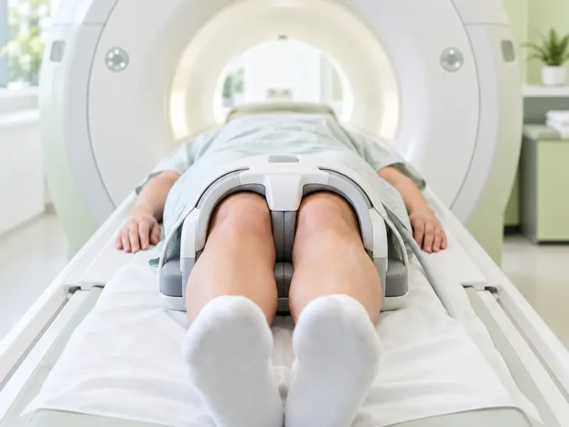
Before any joint treatment, a doctor needs to see what is happening inside the joint. X-ray, MRI and ultrasound each answer different questions, and the right choice depends on the joint and the problem. This article explains what each scan shows, which one usually fits which situation, and how to prepare and send your scans for a review from abroad.
The main points in 30 seconds
- X-ray shows bones and joint space and is often the first scan for joint wear.
- MRI shows cartilage, meniscus, ligaments, tendons and bone marrow in detail.
- Ultrasound shows tendons and fluid and guides the needle during PRP and SVF.
- Most people need one or two scans; the doctor chooses them by joint and symptoms.
- Send images, reports, dates and a short symptom description for a free online review.
Which scans you need before joint treatment
A scan is not a formality. It shows the stage of the condition, rules out problems that need a different approach and helps the doctor decide whether an injection treatment makes sense at all. The same knee pain can come from worn cartilage, a torn meniscus, inflamed tendons or fluid in the joint, and each of these looks different on each type of scan.
In practice, most people need one or two scans, not all three. An X-ray is often the starting point for joint wear, MRI is added when soft tissues or early changes matter, and ultrasound is used to look at tendons and fluid and to guide the needle during the procedure itself.
X-ray: bones, joint space and stage
An X-ray shows bones well and soft tissues poorly. Cartilage is not visible directly, but its thickness can be judged from the gap between the bones, called the joint space. When cartilage wears, this gap narrows and small bony spurs appear at the edges of the joint.
- What it shows: joint space narrowing, bony spurs, changes in the bone under the cartilage, deformity, old fractures.
- What it misses: early cartilage damage, meniscus and ligament tears, tendons, inflammation in soft tissues.
- Useful detail: for the knee and hip, an X-ray taken while standing shows the real load on the joint and makes narrowing easier to see.
X-rays use a small dose of radiation. If you are or may be pregnant, tell the staff before the scan.

MRI: cartilage, meniscus, ligaments and bone marrow
MRI uses a strong magnetic field and radio waves instead of radiation. It gives the most detailed picture of the soft structures inside and around a joint.
- What it shows: cartilage thinning and defects, meniscus tears, ligament and tendon damage, swelling in the bone marrow, fluid and cysts.
- When it is especially useful: when the X-ray looks almost normal but pain persists, when a meniscus or tendon tear is suspected, or when the doctor needs to plan treatment precisely.
- Before the scan: tell the staff about a pacemaker, metal implants, fragments of metal in the body or pregnancy. The scan is painless but noisy and takes some time, and you need to lie still.
Ultrasound: soft tissue, fluid and guided injections
Ultrasound uses sound waves and has no radiation. It shows tendons, ligaments near the surface, fluid in the joint and signs of inflammation. A big advantage is that it works in real time: the doctor can watch a tendon while you move the joint.
Ultrasound does not see through bone, so it cannot show the deep parts of a joint or the cartilage hidden between bones. That is why it rarely replaces an X-ray or MRI for staging joint wear.
Ultrasound also plays a second role: it guides the needle during an injection. Placing PRP or SVF precisely into the joint under ultrasound control is part of how the procedure is done on our joint treatment page.
Which scan fits which joint and problem
Knee wear, pain on stairs
- Usually most informative
- Standing X-ray
- Often added
- MRI for cartilage and meniscus
Suspected meniscus tear
- Usually most informative
- MRI
- Often added
- X-ray to assess wear
Hip pain, groin pain
- Usually most informative
- X-ray of the pelvis and hip
- Often added
- MRI if the X-ray is unclear
Shoulder pain when lifting the arm
- Usually most informative
- Ultrasound or MRI of the tendons
- Often added
- X-ray for joint wear
Swollen joint, fluid
- Usually most informative
- Ultrasound
- Often added
- Blood tests if inflammatory arthritis is suspected
Ankle pain after old sprains
- Usually most informative
- X-ray
- Often added
- MRI for cartilage and ligaments
This is a general guide. The doctor decides which scans are needed after hearing your symptoms and looking at what you already have.
How to prepare and send your scans from abroad
A remote review works well when the doctor gets the full picture. Before sending, gather:
- The images themselves, ideally in the original DICOM format on a disc or as a download link, not only photos of the film.
- The radiologist's written report for each scan. If it is not in English, send it anyway; a translation helps but is not required for a first look.
- The dates of the scans and which side was examined.
- A short description of your symptoms: where it hurts, since when, what makes it worse, previous injuries, injections or operations.
How recent should the scans be? As a rule, images from the last several months are the most useful. If the pain has changed a lot, after a new injury or when the scans are old, the doctor may ask for new ones before making a plan.

Getting a personal plan at City Medical Centre
City Medical Centre works with international patients and treats joints without surgery: with PRP from your own blood and SVF cell therapy from your own adipose tissue, injected under ultrasound guidance. Surgical options such as arthroscopy or joint replacement are not performed at the clinic; if your scans show that one of them is the better choice, the doctor will tell you so openly.
The first consultation and the review of your scans are free and can be done online: send your X-rays, MRI or ultrasound with the reports and a short description of your symptoms. If something is missing, the doctor tells you which scan is needed. If PRP or SVF suits your case, the doctor explains the plan and names the cost, which is then fixed in the contract. A personal coordinator stays with you from the first message to the follow-up visits, and scans and questions can be sent by Telegram. Read more on the joint treatment page.
Sources
Frequently asked questions
Do I need an MRI before PRP injections?
Not always. For joint wear, a recent X-ray is often enough to judge the stage. MRI is added when early cartilage damage, a meniscus or tendon tear, or bone marrow changes need to be checked. The doctor decides after looking at your symptoms and the scans you already have.
Is an X-ray enough to diagnose osteoarthritis?
In many cases an X-ray together with an examination is enough to confirm osteoarthritis and estimate its stage. It does not show early cartilage changes or soft tissues, so when the picture is unclear or pain does not match the X-ray, an MRI may be recommended.
What does a joint ultrasound show?
It shows tendons, ligaments close to the surface, fluid in the joint and signs of inflammation, and it lets the doctor watch structures while you move. It does not see through bone, so deep parts of the joint and cartilage between bones are better assessed with X-ray or MRI.
Can I send my scans for review before travelling?
Yes. Send the images, preferably as DICOM files or a download link, together with the radiologist's reports, the dates of the scans and a short description of your symptoms. The doctor reviews them, says whether anything is missing and whether PRP or SVF may suit you.
How recent should my joint scans be?
Scans from the last several months are usually the most helpful. If there has been a new injury, a clear change in symptoms, or the scans are old, the doctor may ask for new ones so that the plan is based on the current state of the joint.
Have a question about your case? Get a free consultation
Message a coordinator on Telegram: we reply right in the chat, and you can send photos and questions there too.
- The first consultation is free, online consultations are available
- The doctor names the cost after the assessment, and it is fixed in the contract
- A personal coordinator guides you from the first message to recovery
Message our coordinator on Telegram
We reply right in the chat — you can send photos
Message us on TelegramBy sending a message, you agree to the privacy policy
Read also
 Joint treatment
Shoulder joint pain: treatment without surgery and the signs that an operation is needed
Most shoulder pain settles without an operation. Learn the common causes, which scans help, how physiotherapy, PRP and SVF are used and when surgery is needed.
 Joint treatment
SVF cell therapy for joints: how it works and who it suits
SVF cell therapy for joints explained: where the cells come from, which joints it helps, the procedure, recovery, SVF vs PRP and contraindications.
 Rhinoplasty
Septoplasty recovery: what to expect and how to heal well
How recovery after septum surgery usually goes: the first days, splints and crusts, when breathing improves, what to avoid and how to support healing.