


























Joint treatment6 min read
Avascular necrosis of the hip: what can be done in the early stages
Avascular necrosis of the hip develops when part of the head of the thigh bone loses its blood supply. In the early stages, before the bone collapses, there is a real chance to protect the joint. This article explains why the condition develops, which signs to watch for, why MRI is the key scan, what can be done at each stage and when injections are no longer enough.
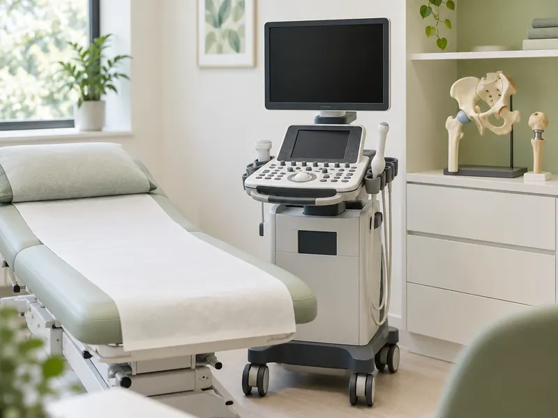
The main points in 30 seconds
- Avascular necrosis is bone death in the femoral head caused by poor blood supply.
- The key goal is to act before the femoral head collapses and loses its shape.
- Early changes are seen on MRI; an X-ray is often normal at first.
- SVF and PRP are used in early stages as part of a plan, not as a guarantee.
- After collapse, hip replacement is often discussed with a surgeon.
Can early avascular necrosis of the hip be treated?
Yes, and timing matters more than in most joint conditions. Avascular necrosis, also called osteonecrosis, is a condition in which bone tissue in the femoral head dies because it does not get enough blood. At first the shape of the bone is preserved. Later, the weakened area can crack and flatten, and the hip joint then wears quickly.
The goal of early treatment is to keep the femoral head round for as long as possible. Once it has collapsed, options narrow and joint replacement becomes more likely. That is why hip or groin pain without a clear cause, especially with known risk factors, deserves prompt attention.
What happens to the bone and why
The head of the femur has a limited blood supply. When the small vessels are damaged or blocked, bone cells in the affected area stop receiving oxygen and die. The body tries to repair the area, but the repair process itself can temporarily weaken the bone under the cartilage.
Known causes and risk factors include:
- hip fracture or dislocation that damaged the blood vessels;
- long-term treatment with high doses of steroid medicines, prescribed for other conditions;
- heavy alcohol use over many years;
- some blood disorders, such as sickle cell disease;
- certain other conditions and treatments, including radiotherapy and decompression illness in divers.
In some people no clear cause is found. The condition often affects both hips, so the second side is usually checked as well.
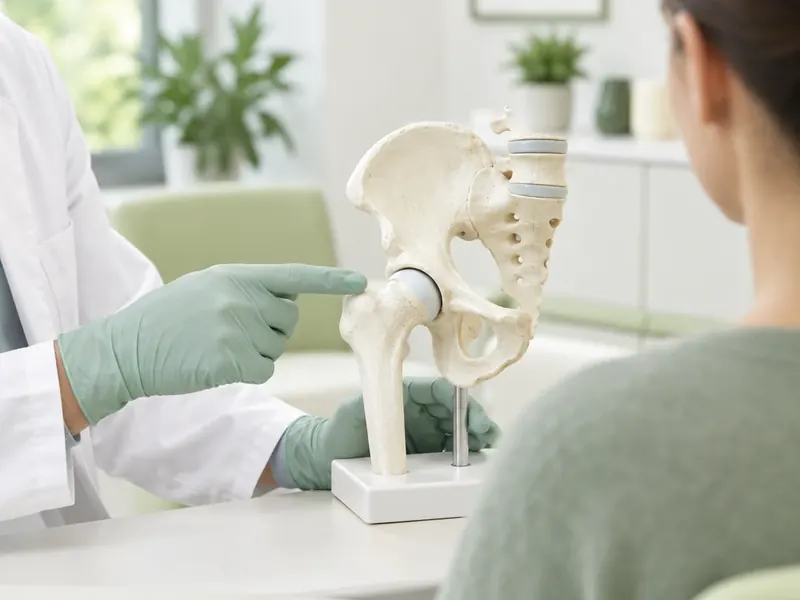
Early symptoms and risk factors
In the very first stage there may be no symptoms at all, and changes are found by chance. When pain appears, it usually:
- is felt in the groin, sometimes in the buttock or thigh, and can spread towards the knee;
- gets worse with walking and standing and eases with rest at first;
- later also appears at rest and at night;
- comes with stiffness, especially when turning the leg inwards;
- may lead to a limp.
If you have one of the risk factors above and develop hip or groin pain, tell your doctor about it directly. This helps them think of avascular necrosis early rather than treating the pain as simple strain.
Why MRI is key to early diagnosis
In the early stage an X-ray is often completely normal, because the shape of the bone has not yet changed. MRI shows swelling in the bone marrow and the typical outline of the affected area weeks or months before an X-ray does. It also shows how large the area is and where it lies, which affects the outlook.
X-rays are still useful: they show whether the femoral head has started to flatten and whether the joint itself is worn. In practice the doctor looks at both. If your hip MRI is older than a few months or was done before the pain changed, a new one may be needed. More on the joints we treat is on our joint treatment page.
Stages and what can be done at each
Doctors use several classification systems. A simplified picture looks like this:
Early, before collapse
- What is happening
- Changes visible on MRI, X-ray normal or nearly normal
- Typical approach
- Reducing load, treating the cause, regenerative injections in suitable cases, joint-preserving surgery
Beginning of collapse
- What is happening
- A thin crack under the cartilage, first flattening of the femoral head
- Typical approach
- Joint-preserving options still considered case by case
Collapse
- What is happening
- The femoral head has lost its round shape
- Typical approach
- Pain management; replacement is often discussed
Advanced arthrosis
- What is happening
- Both joint surfaces are worn
- Typical approach
- Hip replacement is usually recommended
In the early stages, general measures matter too: reducing weight-bearing on the hip for a period, often with crutches, stopping alcohol, and reviewing steroid treatment together with the doctor who prescribed it. Medicines are chosen by the doctor individually. Surgical options such as core decompression, in which channels are drilled into the femoral head to relieve pressure, are general information here; whether they suit you is decided at a consultation with a surgeon.
Regenerative options: SVF and PRP in early stages
SVF is the stromal vascular fraction, a mix of cells isolated from a small sample of your own adipose tissue. PRP is platelet-rich plasma prepared from your own blood. Combined and injected into the hip joint under ultrasound guidance, they are used to reduce pain and inflammation and to create better conditions for the tissues.
These methods make most sense before the femoral head collapses, when there is still a joint to protect. They are part of a plan together with load management and treatment of the cause, not a replacement for them. The response differs from person to person, and no one can promise that the process will stop. The doctor decides after reviewing a recent MRI and X-ray whether SVF or PRP is reasonable in your case.
When injections are not enough
Injections are unlikely to help enough when:
- the femoral head has already collapsed and changed shape;
- the affected area is very large;
- the joint surfaces are worn and pain at rest and at night is severe;
- walking is seriously limited despite treatment.
In these situations hip replacement is usually discussed with an orthopaedic surgeon. It is general information here; the decision depends on your scans, age and activity. If injections are not the right choice for you, the doctor will say so rather than offer a procedure that is unlikely to help.
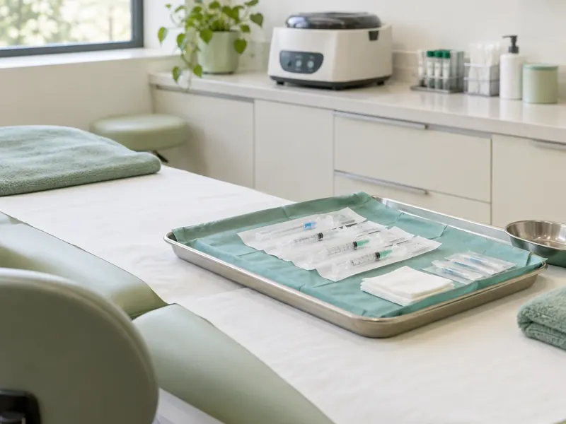
Getting a personal plan at City Medical Centre
City Medical Centre works with international patients and treats joints, including early avascular necrosis of the hip, without surgery: with SVF cell therapy from your own adipose tissue and PRP from your own blood, injected under ultrasound guidance. The clinic does not perform core decompression or hip replacement; if your scans show that surgery is the better option, the doctor will tell you so openly. The first consultation and the review of your MRI and X-rays are free and can be done online.
If SVF or PRP suits your stage, the doctor explains the plan and names the cost, which is then fixed in the contract. A personal coordinator stays with you from the first message to the follow-up visits, and scans and questions can be sent by Telegram. Read more on the joint treatment page.
Sources
Frequently asked questions
Can avascular necrosis of the hip be reversed?
Bone that has already died does not come back, but in the early stages, before the femoral head collapses, the process can sometimes be slowed and the joint preserved for a long time. The earlier the diagnosis, the more options there are. How the condition develops differs from person to person.
Does SVF therapy help early hip necrosis?
SVF combined with PRP is used in early stages to reduce pain and inflammation and support the tissues, as part of a plan that includes load management and treating the cause. It makes most sense before collapse. Results vary, and the doctor decides after reviewing a recent MRI whether it suits you.
What are the first signs of avascular necrosis?
Often pain in the groin, sometimes in the buttock or thigh, that gets worse with walking and standing. Later it appears at rest and at night, and turning the leg inwards becomes stiff and painful. In the very first stage there may be no symptoms at all.
Which scan detects avascular necrosis early?
MRI. In the early stage an X-ray is often normal, while MRI already shows the affected area and swelling in the bone marrow. X-rays are then used to check whether the femoral head has started to flatten and how worn the joint is.
When is hip replacement needed for necrosis?
It is usually discussed when the femoral head has collapsed, the joint surfaces are worn, and pain and limited walking persist despite treatment. The decision is made by an orthopaedic surgeon based on your scans, age and activity.
Have a question about your case? Get a free consultation
Message a coordinator on Telegram: we reply right in the chat, and you can send photos and questions there too.
- The first consultation is free, online consultations are available
- The doctor names the cost after the assessment, and it is fixed in the contract
- A personal coordinator guides you from the first message to recovery
Message our coordinator on Telegram
We reply right in the chat — you can send photos
Message us on TelegramBy sending a message, you agree to the privacy policy
Read also
 Joint treatment
Elbow joint pain: arthritis, arthrosis and treatment without surgery
Elbow pain can come from joint wear, inflammation or overloaded tendons. Learn the causes, symptoms, diagnosis and how rehab, PRP and SVF are used without surgery.
 Joint treatment
PRP and SVF joint treatment cost: what you pay for and how to compare offers
What shapes the cost of PRP and SVF joint injections: the method, number of joints and sessions, scans, follow-up, and how to compare offers fairly.
 Bariatric surgery
Bariatric surgery cost: what affects the price and what should be included
What really drives the price of bariatric surgery: the type of operation, tests, hospital stay, aftercare and vitamins, and how to get a fixed quote.